Ferroptossis _ an Iron and lipid induced cell death

Iron-dependent lipid peroxidation is characterized by unique biochemical and morphological changes which are distinct from other forms of cell death apoptosis, necrosis, and autophagy
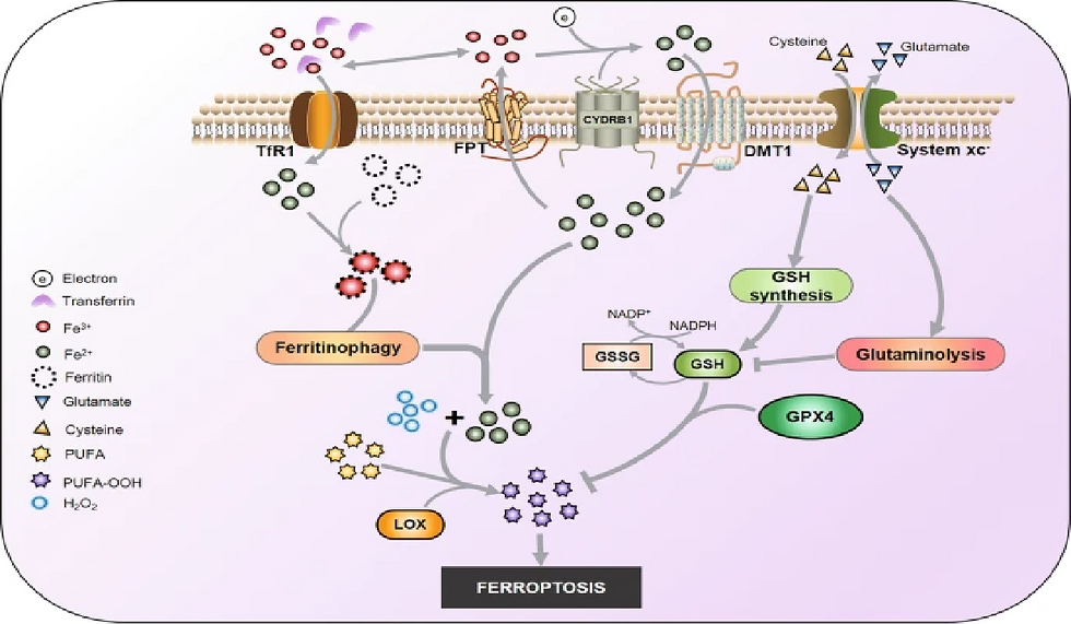
Lipid peroxidation coupled with the Fenton reaction induces ferroptosis
Lipid peroxidation occurs through two mechanisms :
Nonradical chain reaction (Fenton reaction)
Enzymatic LOXS

IREB2: It is a major regulator of iron metabolism

When cells are iron deficient, IRPs bind to 5′ IREs in ferritin and ferroportin mRNAs with high affinity to repress translation, and to 3′ IREs in TfR1 mRNA to block its degradation
When iron is in excess, IRPs do not bind to IREs, increasing the synthesis of ferritin and ferroportin, while promoting the degradation of TfR1 mRNA
The role of mitochondria
The role of mitochondria in ferroptosis is context-dependent; if the activity of the glutathione-dependent peroxidase, GPX4, is inhibited, cells undergo ferroptosis independent of mitochondrial function.
So, it is important to check by which factor ferroptosis is induced
Characteristic features of ferroptosis which is unique to ferroptosis
Increase in lipid peroxides (C11 BODIPY)
No change in mitosox
Morphological changes in mitochondrial cristae
No caspase activation or no formation of membrane-bound vesicles (like in autophagy)
Potential genes /Markers for ferroptosis (multiple genes associated with iron uptake, lipotoxicity, and antioxidation responses)
Lipid peroxidation :
1. ALOXs: Arachidonate lipoxygenases
Catalyze peroxidation of AAs (PUFAs)
2. ACSL4- Acyl-CoA synthetase long-chain family member 4
3. LPCAT- lysophosphatidylcholine acyltransferase 3
4. CS- Cysteine synthetase
5. GPX4: Glutathione peroxidase 4
Reduces LOOHs in membrane phospholipids to suppress ferroptosis
Iron metabolism :
IREB2: It is a major regulator of iron metabolism
Compounds that induce ferroptosis
Inhibition of system Xc-
Prevents cystine import, causes GSH depletion and loss of GPX4 activity
Eg: Erastin
Direct inhibition of GPX4
Direct inhibition of GPX4 Covalently interacts with GPX4 and inhibits its enzymatic activity
Eg: RSL3, ML162, DPI compounds
Induction of lipid peroxidation
Oxidizes iron, drives lipid peroxidation and indirect inactivation of GPX4
Eg: FINO2
Inhibitors of ferroptosis
Class 1: Iron chelators
Deplete iron and prevent iron-dependent lipid peroxidation
Eg: Deferoxamine, ciclopirox, deferiprone
Class 2: Lipophilic antioxidants
Prevent lipid peroxidation
Eg: Vitamin E, BHT, Ferrostatin -1, liproxstatin-1, XJB-5- 131, CoQ10
Class 3: D-PUFAs
Prevents initiation and propagation of lipid peroxidation
Eg: D4-arachidonic acid, D10-docosahexaenoic acid
Class 4: LOX inhibitors
Inactivate LOX and block LOX-induced lipid peroxidation
Eg: DC, baicalein, PD-146176, AA-861, zileuton
Ferroptosis, a regulated form of cell death driven by iron overload, glutathione depletion, and lipid peroxidation, is triggered by several pathological or stress-induced factors, including excess iron, inhibited GPX4 activity, and elevated ROS levels. Studying ferroptosis is crucial because it enhances our understanding of disease mechanisms, particularly in cancer and neurodegenerative disorders, and reveals potential therapeutic targets for drug development. Identifying ferroptosis-specific biomarkers can aid in early disease detection and monitoring. At the same time, insights gained from this research can lead to novel treatments that induce ferroptosis in cancer cells or prevent it in conditions where excessive cell death is harmful.
References
1. BioTek. (n.d.). *An introduction to reactive oxygen species: Measurement of ROS in cells*. Retrieved from https://www.biotek.com/resources/white-papers/an-introduction-to-reactive-oxygen-species-measurement-of-ros-in-cells/
2. Tang, D., Chen, X., Kang, R., & Kroemer, G. (2012). *The molecular machinery of ferroptosis*. Cell, 153(3), 489-495. https://doi.org/10.1016/j.cell.2012.03.042
3. Dixon, S. J., Lemberg, K. M., Lamprecht, M. R., et al. (2012). *Ferroptosis: An iron-dependent form of nonapoptotic cell death*. Proceedings of the National Academy of Sciences, 109(43), 16648-16653. https://doi.org/10.1073/pnas.0912074106
4. Sun, X., Niu, X., & Zhang, J. (2020). *The role of erastin in ferroptosis and its prospects in cancer therapy*. Frontiers in Pharmacology, 11, 1371. https://doi.org/10.3389/fphar.2018.01371
5. Chien, W.-C., & Liao, J.-W. (2019). *The role of acyl-CoA synthetase long-chain family member 4 (ACSL4) in ferroptosis and cancer*. eLife, 8, e47733. https://elifesciences.org/articles/47733
6. Yagoda, N., Von Rechenberg, M., & Zaganjor, I. (2007). *RAS–RAF–MEK-dependent oxidative cell death involving voltage-dependent anion channels*. Nature, 447(7146), 864-868. https://doi.org/10.1038/nature05827
7. Feng, H., & Stockwell, B. R. (2018). *Unsolved mysteries: How does lipid peroxidation cause ferroptosis?* PLOS Biology, 16(5), e2006203. https://doi.org/10.1371/journal.pbio.2006203
8. Theodossiou, S., & Georgopoulos, A. (2004). *Oxidative stress and retinal vascular changes*. Investigative Ophthalmology & Visual Science, 45(6), 2054-2061. https://doi.org/10.1167/iovs.03-1355


Comments